-
Anatomical models
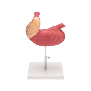
HUMAN STOMACH
This model shows the the major regions of the stomach and also the inside of the stomach. Lengthwise folds of the stomach lining, called rugae, allow the stomach to expand and partially digest food. Large size, showing external details, mounted on stand, with key card.
BG12170 -
Anatomical models
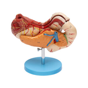
HUMAN STOMACH PANCREAS & DUODENUM
The stomach wall is illustrated in the model, showcasing its various layers.
The model provides a visual representation of the lower esophagus, along with the associated blood vessels. Additionally, it displays sectioned portions of the duodenum and pancreas, revealing the presence of large ducts leading up to its opening. Divided into three parts, the model is mounted on a plastic stand and comes with a key card.BG12172 -
Anatomical models
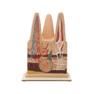
INTESTINAL VILLI MODEL-100 TIMES ENLARGED
This highly detailed anatomical model of intestinal villi is enlarged 100 times to provide an in-depth view of its complex structure. The model features one entire villus, one longitudinally sectioned villus displaying the arterioles and venules, and one cut villus that reveals the lymphatic vessels. Additionally, the model includes an enlarged representation of a longitudinal section of Lieberkuhn’s crypt, offering a comprehensive understanding of the micro anatomy of the intestinal lining. This model is an invaluable educational tool for students and professionals, providing clear visualization of the microscopic structures involved in nutrient absorption.
BG12173 -
Anatomical models
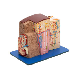
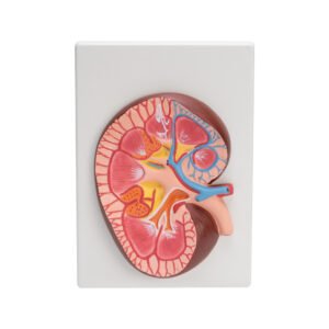
KIDNEY MICROSTRUCTURE
A complete 3-D model shows 5 side view of following structures with great details : kidney in section , parenchyma of Kidney and Nephron, renal corpuscle and associated structures – Loop of Henle and associated structures.
Mounted on plastic base with key card.BG12174 -
Anatomical models
HUMAN KIDNEY WITH ADRENAL GLAND MODEL
This detailed anatomical model of the human kidney with an adrenal gland is dissectable into two parts to provide an in-depth view of its structure. The model showcases the kidney with the adrenal gland, including renal and adrenal vessels and the upper portion of the ureter. The front half of the kidney is removable, revealing the cortex, medulla, and vessels, as well as the renal pelvis. Mounted on a sturdy stand for stability, this model is designed for easy examination and study. It comes with a key card that labels and explains each component, making it an invaluable educational tool for students, educators, and medical professionals.
BG12177 -
Anatomical models
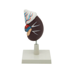
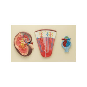
RENAL STRUCTURE AND FUNCTION MODELS
This set of three anatomical models, mounted on a single board, provides a detailed look at kidney structure and function. The Kidney Section is enlarged 3 times to show the cortex and medulla, highlighting the overall architecture and key anatomical features. The Nephrons and Blood Vessels model, enlarged 120 times, illustrates sections through the renal cortex and medulla, focusing on the nephrons and their associated blood vessels, providing a clear depiction of the intricate network involved in blood filtration and urine formation. The Malpighian Corpuscle model, enlarged 700 times, showcases an open corpuscle with the glomerulus and Bowman’s capsule, helping to understand the initial stages of blood filtration in the kidney. Mounted on a stable base with a key card for easy identification, this model set is ideal for educational use by students, educators, and medical professionals.
BG12177A -
Anatomical models
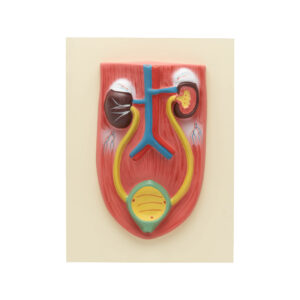
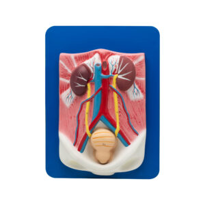
KIDNEY WITH BLADDER MODEL
This anatomical model, presented at natural size, provides a comprehensive view of the kidney, ureters, adrenal glands, and bladder. The large abdominal right kidney is sectioned to reveal all anatomical details, offering an in-depth understanding of the urinary system. Mounted on a sturdy base for stability and ease of display, this model is an excellent educational tool for students, educators, and medical professionals, facilitating a detailed study of the urinary and adrenal systems.
BG12177B -
Anatomical models
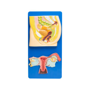
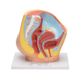
HUMAN REPRODUCTIVE SYSTEM FEMALE
Upper half model shows both ovaries, left ovary in section, fallopian tubes with fimbriae, uterus, cut open vagina to show vaginal folds and cervix, bladder with cut open urethra, labia and rectum. Lower half model shows adnexa of uterus.
Mounted on wooden base with key card.BG12178 -
-
Anatomical models
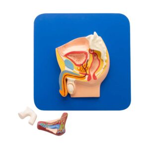
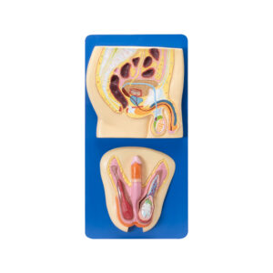
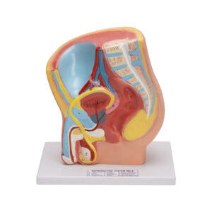
HUMAN REPRODUCTIVE SYSTEM MALE
The upper half of the model provides a detailed median section of the human male reproductive system, showcasing internal organs such as the epididymis, vas deferens, ejaculatory ducts, urethra, seminal vesicles, prostate gland’s external structure, penis, scrotum, and testicles. The entire model is securely mounted on a wooden board and includes an informative key card.
BG12184 -
Anatomical models
HUMAN URINARY ORGANS MODEL
This natural-size anatomical model of the human urinary organs is dissectable into three parts, providing a detailed view of the kidneys, ureters, adrenal glands, bladder with prostate, and major blood vessels. The right kidney is sectioned to reveal its internal structure, and the bladder and prostate are removable for closer examination. Mounted on a sturdy base, this model comes with a key card that labels and explains each part, making it an invaluable educational tool for students, educators, and medical professionals to study the urinary system in detail.
BG12186 -
Anatomical models
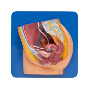
FEMALE PELVIS
Female Pelvis Model showcases the median section and includes removable inner organs. It depicts the external and internal genital organs, along with the pelvic muscles, muscles of the pelvic floor, and the intricate network of nerves and vessels.
Mounted on plastic base with key card.BG12190 -
Anatomical models
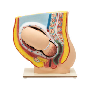
PREGNANCY PELVIS WITH BABY MODEL
This anatomical model shows a median section through the female pelvis at the 9th month of pregnancy, complete with a removable fetus. It allows for the study of the normal position of the child before birth. Mounted on a sturdy base, the model provides a realistic and detailed view of the pelvis and fetus, making it an invaluable educational tool for students, educators, and medical professionals. The model comes with a key card that labels and explains each part, enhancing the learning experience.
BG12201 -
Anatomical models
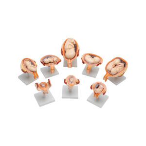
Embryonic Development Model Set (Period of Gestation)
The Embryonic and Fetal Development Model set provides a sequential representation of prenatal growth stages from the 1st to the 7th month. Each model is crafted with detailed anatomical accuracy, illustrating key developmental milestones, including early organ formation, limb growth, and positioning within the womb. 1st Month Embryo, features the foundational structures of the neural and heart tubes, early limb buds, and yolk sac that provides initial nutrients. 2nd Month Embryo, shows early formation of facial features (eyes, nose, mouth), limb growth, and the beginning of organ differentiation. 3rd Month Embryo, presents fully formed limbs, a more defined face, and early reflexive movements. 4th Month Embryo, includes fine hair (lanugo), hardened bones, and distinct genitalia, indicating further development and gender differentiation. 5th Month Fetus (Normal Position) depicts active movement, skin covered with protective vernix. 5th Month Fetus (Transverse Lie) represents the fetus positioned horizontally across the uterus, a temporary position often monitored for later adjustment. 7th Month Fetus shows open eyes, developing fat deposits, and strong reflexes for sucking and swallowing, indicating advanced readiness for birth.
BG12201A