Biology
-
Zoological models
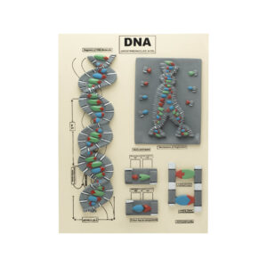
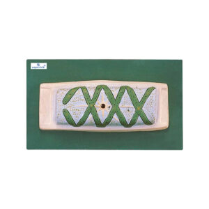
DNA (DEOXYRIBONUCLEIC ACID) MODEL
This DNA model provides a comprehensive representation of the DNA molecule, highlighting its structure, replication mechanism, and molecular details:
Double Helix Structure: Illustrates the double helix coiling and reforming, showcasing the spiral shape of DNA.
Nucleotides: Displays the nucleotide components (adenine, thymine, cytosine, and guanine) and their arrangement within the DNA strands. Mechanism of Replication: Shows how DNA replicates, emphasizing the complementary base pairing and enzymatic processes involved. Structural Formula: Includes the structural formula to depict the molecular shapes, bonds, and angles, emphasizing the chemical composition and configuration of DNA. The complete model is meticulously arranged on a board, fully labeled with detailed annotations. This educational tool provides a clear understanding of DNA’s molecular structure and function, making it ideal for students, educators, and researchers studying genetics, molecular biology, and biochemistry.BG12688A -
-
Zoological models
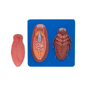
COCKROACH DISSECTED MODEL
One model intricately displays the cockroach’s internal anatomy, highlighting the alimentary canal, salivary glands, reproductive organs, and nervous system. Another model focuses on the cockroach’s ventral anatomy, featuring detachable components such as mouthparts, wings, and appendages, accompanied by an informative key card for detailed study.
BG12692 -
Zoological models
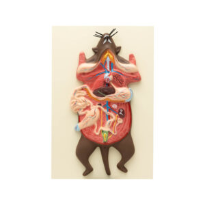
RAT DISSECTION
Dissection of the domestic rat showing the internal viscera, chief systems, alimentary canal and its various parts, with glands, circulatory, respiratory, excretory and the female genital organs. The head is longitudinally cut to show the brain, buccal and nasal cavities, mounted on board, with key card.
BG12694 -
Botanical models
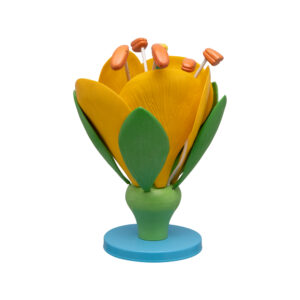
TYPICAL FLOWER
Large-scale model depicting a typical flower, featuring detachable parts including the ovary housing a single ovule, sterile whorls representing the calyx and corolla, and fertile whorls representing the androecium and gynoecium. Mounted on a base and accompanied by a key card for reference.
BG12810 -
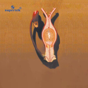
Botanical models
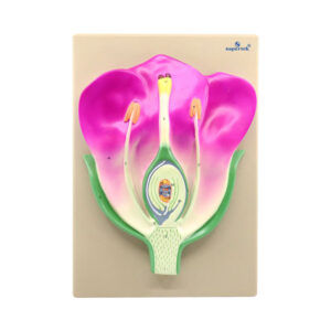
TYPICAL FLOWER L.S.
A detailed model illustrating the longitudinal section of a flower, highlighting its internal structure and various components. This includes the pistil, stamen, and sepal, as well as the pedicel, thalamus, calyx (sepals), corolla (petals), nectary, filament, anther lobe, ovary, ovule, style, stigma, pollen grain, pollen tube, first male gamete, second male gamete, female gamete (oosphere), synergids, secondary nuclei, antipodals, embryo sac, and nucellus. The model is securely mounted on a board and accompanied by a key card for easy reference.
BG12812 -
Botanical models
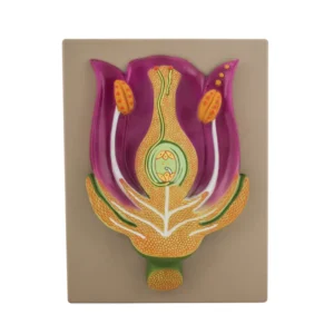
MODEL TYPICAL FLOWER V.S.
Delve into the inner workings of the stem, sepal, petal, stamen, and pistil as each component is revealed. This interactive model allows students to trace the transmission of male gametes to female gametes within the ovary, enhancing their understanding of pollination and fertilization processes. Mounted on a base and accompanied by a key card for reference.
BG12812A -
Botanical models
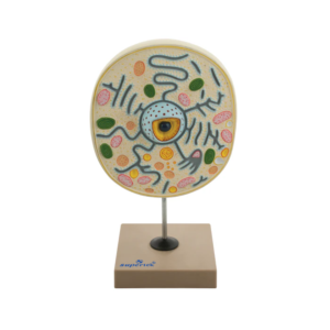
TYPICAL PLANT CELL
Intricate details of plant cell structure with this magnified model, resembling views under an electron microscope. With sections revealing ectoplast, endoplast, tonoplast, vacuoles, nuclear structure, plastids, and mitochondria, this model provides comprehensive insight. Mounted on a base for stability and accompanied by a key card for easy reference.
BG12820 -
Botanical models
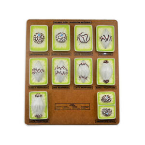
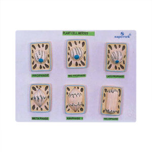
PLANT MITOSIS
Presenting a set of 10 models illustrating the complete process of karyokinesis and cytokinesis, from the metabolic cell to the formation of two daughter nuclei. Each model identifies the key components of cell phases during mitosis, highlighting changes cells undergo throughout the stages. Featuring detailed representations of the nucleus, centrioles, centrosome, chromatin, chromosomes, spindle, and aster, all stages are mounted on a board for easy display and accompanied by a key card for reference.
BG12830 -
Botanical models
MITOSIS PHASES DEMONSTRATION MODEL
Hand-painted chromosomes in vibrant colors depict chromosomal movement. Ideal for classroom demonstrations, painted with ecofriendly paint and designed by subject matter experts. Mounted on a base and accompanied by a key card for reference.
BG12832A -
Botanical models
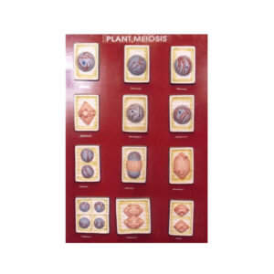
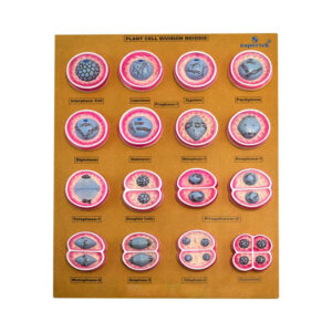
PLANT CELL DIVISION MEIOSIS
Explore the latest understanding of chromosome changes with this set of 15 newly designed models. From the resting nucleus to the formation of four daughter cells, each phase is depicted and mounted on a board. Phases and details include interphase, prophase-1 (leptotene, zygotene, pachytene, and diplotene), diakinesis, metaphase-1, anaphase-1, telophase-1, daughter cells, prophase-2, metaphase-2, anaphase-2, telophase-2, and gametes.
BG12835 -
-
Botanical models
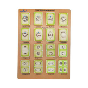
PLANT CELL DIVISION MASTER MODEL SET
Presenting a set of 16 meticulously hand-painted plant cell division models mounted on a board, showcasing intricate detail. These models represent an entirely new design based on recent concepts of chromosome changes, depicting the process from the resting nucleus to the formation of 4 daughter cells. Each model is numbered for easy reference with an english key card.
BG12842A -
Botanical models
MEIOSIS PHASES EXPLORATION MODEL
This model offers a detailed rendering of meiosis, featuring tough fiber glass molded cell models. Phases include inter phase, prophase- 1(leptotene, zygotene, pachytene, and diplotene), diakinesis, metaphase-1, anaphase-1, telophase-1, daughter cells, prophase-2, metaphase-2, anaphase-2, telophase-2, and gametes.
Mounted on a base and accompanied by a key card for reference.BG12842B -
Botanical models
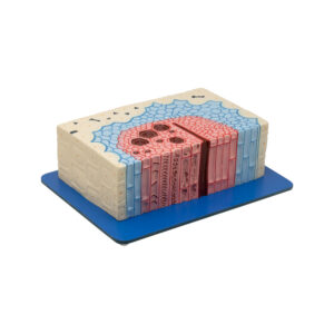
PLANT TISSUE CAMBIUM MODEL
Intricate structure of plant tissue with this model, showcasing cambium between xylem and phloem. A single strip of phloem with a single cambium is depicted, featuring both open and closed bundles. Enlarged approximately 550 times, this detailed model is presented in one piece on a base, accompanied by a key card for reference.
BG12846 -
Botanical models
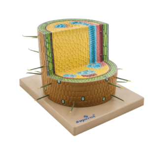
DICOTYLEDONOUS STEM ANATOMY MODEL
Explore the tissue structure of a garden bean’s dicotyledonous stem with this model, depicting a cross-section magnified 200 times. Mounted on a base for stability, each component is numbered for easy identification with an English Key Card.
BG12846A -
Botanical models
FERTILISATION OF ANGIOSPERMS
Discover the polygynous type of reproduction with this model, showcasing male gametes within the embryo sac. The fusion of two sperm cells separately with the egg and central cell is depicted, magnified 300 times for detail. Mounted on a board for easy display and accompanied by a key card for reference.
BG12848