Models
-
-
Models
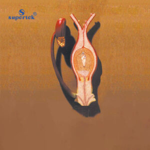
FERTILISATION OF ANGIOSPERMS
Discover the polygynous type of reproduction with this model, showcasing male gametes within the embryo sac. The fusion of two sperm cells separately with the egg and central cell is depicted, magnified 300 times for detail. Mounted on a board for easy display and accompanied by a key card for reference.
BG12848 -
Models
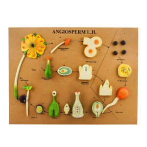
ANGIOSPERM LIFE CYCLE MODEL
It represents the structures involved in the flowering plant’s life cycle, aiding students in visualizing and understanding the process. Clear labelling of life cycle steps and cells enhances comprehension. Perfect for biology classrooms, this model features key structures including flower, stamen, carpel, ovary, pollen tube, ovule, embryo, seed, seedling, and sporophyte.
BG12848A -
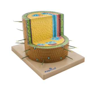
Models
ROOT ANATOMY
This model illustrates the tissue arrangement in plant stems, featuring transverse, longitudinal, radial, and tangential cuts for clear structural details. Monocot roots highlight distinct regions including Epiblema, Cortex, Endodermis, Pericycle, Vascular bundles, and Pith. Mounted on a stand with a key card for reference.
BG12850 -
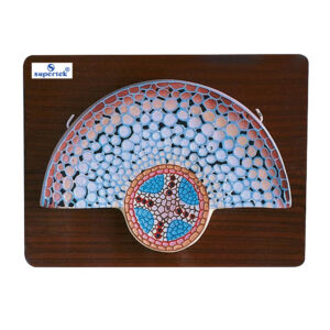
Models
MONOCOT ROOT T.S.
Internal details of Smilax root with this mounted model, featuring two or three layers of sclerenchyma below the epidermis. Hairs are absent, and intercellular spaces are not present. The model showcases an undifferentiated cortex, epidermis, endodermis, pericycle, and pith. Accompanied by a key card for reference.
BG12856 -
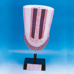
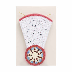
Models
DICOT ROOT T.S.
Internal anatomy of a young dicotyledonous root with this model, showcasing key features such as the taproot system, epidermal cells resembling root hairs, multi-layered cortex, and loose parenchyma cells with intercellular spaces. Mounted on a board for easy display and accompanied by a key card for reference.
BG12858 -
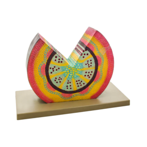
Models
MONOCOT STEM INTERNAL ANATOMY & VASCULATURE
This model showcases the different tissues and vascular bundles of maize, including scattered, closed, and collateral types, in both transverse and longitudinal sections. It allows for detailed examination of internal structures such as large pitted vessels and spiral and annular vessels, revealing cell wall thickenings in longitudinal sections. Highlights parts like the cortex, endodermis, and pericycle. Mounted on a base and accompanied by a key card for reference.
BG12862 -
Models
MODEL MONOCOT STEM
This model showcases the tissue composition, as well as the scattered closed and collateral vascular bundles, in both transverse and longitudinal sections of maize. Highlighting features such as large pitted vessels, spiral, and annular vessels. Mounted on a base and accompanied by an English Key Card.
BG12862A -
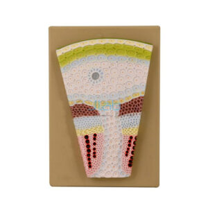
Models
MONOCOT STEM T. S.
Explore the internal structure of a monocot stem from maize with this model, featuring transverse sections of various tissues and vascular bundles. Each section is comprised of a single layer of cells with a thick cuticle. Stomata are present, while multicellular hairs are absent. Mounted on a board for display, accompanied by a key card for easy reference.
BG12872 -
Models
LEAF ANATOMY
Intricate structure of a leaf with this 3-dimensional model, showcasing detailed transverse and longitudinal sections. Featuring loosely packed spongy mesophyll and stomata beneath for gas exchange, this large-size model is mounted on a stand for easy viewing. Accompanied by a key card for reference.
BG12880