Anatomical models
-
Anatomical models
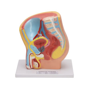
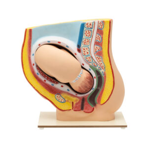
PREGNANCY PELVIS WITH BABY MODEL
This anatomical model shows a median section through the female pelvis at the 9th month of pregnancy, complete with a removable fetus. It allows for the study of the normal position of the child before birth. Mounted on a sturdy base, the model provides a realistic and detailed view of the pelvis and fetus, making it an invaluable educational tool for students, educators, and medical professionals. The model comes with a key card that labels and explains each part, enhancing the learning experience.
BG12201 -
Anatomical models
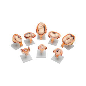
Embryonic Development Model Set (Period of Gestation)
The Embryonic and Fetal Development Model set provides a sequential representation of prenatal growth stages from the 1st to the 7th month. Each model is crafted with detailed anatomical accuracy, illustrating key developmental milestones, including early organ formation, limb growth, and positioning within the womb. 1st Month Embryo, features the foundational structures of the neural and heart tubes, early limb buds, and yolk sac that provides initial nutrients. 2nd Month Embryo, shows early formation of facial features (eyes, nose, mouth), limb growth, and the beginning of organ differentiation. 3rd Month Embryo, presents fully formed limbs, a more defined face, and early reflexive movements. 4th Month Embryo, includes fine hair (lanugo), hardened bones, and distinct genitalia, indicating further development and gender differentiation. 5th Month Fetus (Normal Position) depicts active movement, skin covered with protective vernix. 5th Month Fetus (Transverse Lie) represents the fetus positioned horizontally across the uterus, a temporary position often monitored for later adjustment. 7th Month Fetus shows open eyes, developing fat deposits, and strong reflexes for sucking and swallowing, indicating advanced readiness for birth.
BG12201A -
Anatomical models
HUMAN SPERM MODEL
The sperm model is magnified millions of times, unveiling the internal composition of the gamete: head, mid, tail, centriole, mitochondria, outer dense fibers, and axial filament. It also displays the acrosome, nucleus, centriole, and plasma membrane. This model is mounted on a plastic base with a key card.
BG12202 -
Anatomical models
HUMAN SPINAL CORD MODEL-10 TIMES ENLARGED
This detailed anatomical model of the human spinal cord is enlarged 10 times to provide an enhanced view of its structure and components. The model includes nerve branches, clearly depicting the connections and pathways within the spinal cord. Mounted on a sturdy base for stability and ease of display, it offers a comprehensive understanding of spinal cord anatomy. The model comes complete with a key card that labels and explains each part, making it an excellent educational tool for students, educators, and medical professionals.
BG12203 -
Anatomical models
BONE MICROSTRUCTURE
The models shows 3-D section of a lamellar Bone. The model helps to understand the typical elements of a lamellar bone along with the volkmann & haversian system, spongy & compact parts, endosteum, cortical substance & osteocytes. Model shown various planes in cross and longitudinal section. Mounted on wooden base with key card.
BG12204 -
Anatomical models
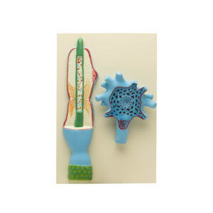
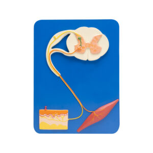
NEURON MODEL – ENLARGED 2500 TIMES
This highly detailed model of a neuron is enlarged approximately 2500 times to vividly display its intricate structure. The model showcases the neuron’s components, including the cell body,
dendrites, axon, and synaptic terminals, with a separate modulated nerve fiber as seen under an electron microscope. Mounted on a sturdy board for stability, this model provides a clear and
comprehensive view of the neuron’s anatomy and its function in the nervous system. It comes complete with a key card that labels and explains each part, making it an invaluable educational tool for students, educators, and medical professionals.BG12205 -
Anatomical models
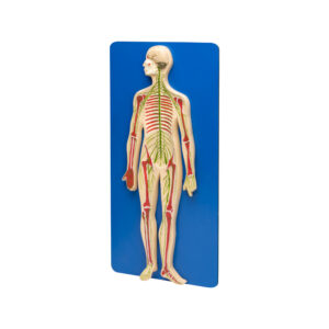
HUMAN NERVOUS SYSTEM
Half life size relief model of human nervous system. Model is a schematic representation of the central and peripheral nervous system, including spinal nerves radiated from central nervous system to other parts of body. Mounted on wooden base with key card.
BG12206 -
Anatomical models
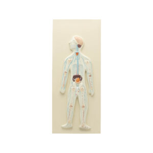
HUMAN NERVOUS SYSTEM MODEL-COMPACT SIZE
This compact anatomical model provides a detailed representation of the human nervous system, including the brain, spinal cord, and nerves in their natural positions within the body. Despite its smaller size, the model accurately depicts the intricate structures and connections of the nervous system. Mounted on a board for stability and ease of display, it offers a comprehensive view of neuroanatomy that is perfect for smaller space sopor table educational settings. The model comes with a key card that labels and explains each component, making it an excellent tool for students, educators, and medical professionals to study and understand the human nervous system.
BG12206A -
-
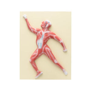
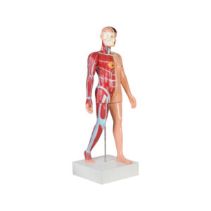
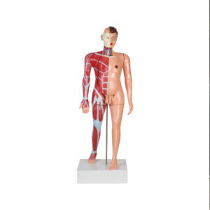
Anatomical models
MUSCLE FIGURE MODEL
This anatomical model, approximately 1/10th natural size, provides a detailed representation of muscle topography. The model displays
the muscles in one piece, offering a comprehensive overview of their arrangement and structure. Mounted on a sturdy base for stability, it
comes with a key card that labels and explains each muscle, making it an excellent educational tool for students, educators, and medical
professionals.BG12226 -
Anatomical models
TORSO FEMALE (FULL SIZE)
This life-sized human female torso model showcases both organs and muscles, providing a comprehensive study of female anatomy. The torso itself is dissected into 20 parts, offering a detailed exploration of internal structures. The model includes removable components, such as the head with a detachable brain, two parts heart, right lung, left lung, liver, stomach, kidney, small and large intestine, two parts female reproductive organs with a fetus, breasts, arms, and legs. Mounted on a movable wooden base with wheels and a center rod & accompanied by a key card for easy reference.
BG12230 -
Anatomical models
TORSO MALE (FULL SIZE)
This life-sized human male torso model showcases both organs and muscles, providing a comprehensive study of male anatomy. The torso is dissected into 20 parts, allowing for an in-depth examination of internal structures. The model includes removable components, such as the head with a detachable brain, two parts for the heart, right lung, left lung, liver, stomach, kidney, small and large intestine, two parts male reproductive organs, chest, arms, and legs.
Mounted on a wooden movable base with wheels and a center rod & accompanied by a key card for easy reference .BG12232 -
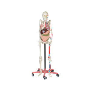
Anatomical models
HUMAN SKELETON WITH MUSCLE & ORGANS
This life-sized human skeleton model is designed for comprehensive demonstrations, featuring hand-painted muscle origins and flexible joints. The skull includes a movable jaw, and the model incorporates detachable parts such as a two-part heart, right lung, left lung, liver, stomach, small intestine, and large intestine. Both arms and legs are movable, allowing for dynamic presentations. The model is securely mounted on a plastic stand and includes a key card for detailed reference.
BG12234