Biology
-
Charts
Botany Series III
These vacuum-formed biological charts are made from heavy-duty plastic, offering deep relief for clear, tactile representation. Weatherproof and washable, they are built to withstand years of
classroom use. With realistic colors and an aesthetically pleasing design, these charts provide valuable assistance in helping students understand biological concepts. They are also specially designed to be helpful for blind students, making learning more accessible.Set of 8
(1) Fertilization
(2) T.S. Dicot leaf
(3) T.S. Monocot leaf
(4) T.S. Dicot root
(5) T.S. Monocot root
(6) T.S. Dicot stem
(7) T.S. Monocot stem
(8) Types of placentationWhile size 25 x 35 cm is available in sets only, other sizes are available singly as well.
BG13155 -
Charts
Botany Series IV
These vacuum-formed biological charts are made from heavy-duty plastic, offering deep relief for clear, tactile representation. Weatherproof and washable, they are built to withstand years of
classroom use. With realistic colors and an aesthetically pleasing design, these charts provide valuable assistance in helping students understand biological concepts. They are also specially designed to be helpful for blind students, making learning more accessible.Set of 8
(1) Plant kingdom
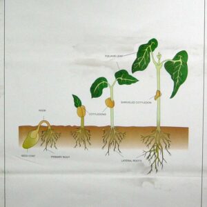
(2) Germination of sunflower and maize
(3) Germination of bean
(4) Yeast and pea
(5) Rhizopus and Mucor
(6) Fern
(7) Insectivorous plants
(8) MalvaceaeWhile size 25 x 35 cm is available in sets only, other sizes are available singly as well.
BG13157 -
Charts
Zoology Series I
These vacuum-formed biological charts are made from heavy-duty plastic, offering deep relief for clear, tactile representation. Weatherproof and washable, they are built to withstand years of
classroom use. With realistic colors and an aesthetically pleasing design, these charts provide valuable assistance in helping students understand biological concepts. They are also specially designed to be helpful for blind students, making learning more accessible.Set of 8
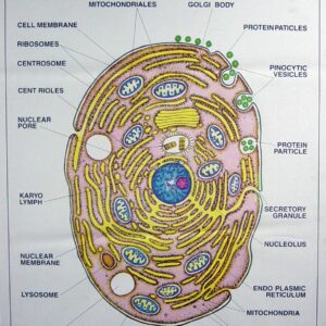
(1) Typical animal cell
(2) Animal mitosis
(3) Hydra
(4) Paramecium
(5) Amoeba proteus
(6) Hookworm
(7) Tapeworm
(8) EarthwormWhile size 25 x 35 cm is available in sets only, other sizes are available singly as well.
BG13159 -
Charts
Zoology Series II
These vacuum-formed biological charts are made from heavy-duty plastic, offering deep relief for clear, tactile representation. Weatherproof and washable, they are built to withstand years of
classroom use. With realistic colors and an aesthetically pleasing design, these charts provide valuable assistance in helping students understand biological concepts. They are also specially designed to be helpful for blind students, making learning more accessible.Set of 8
(1) Euglena
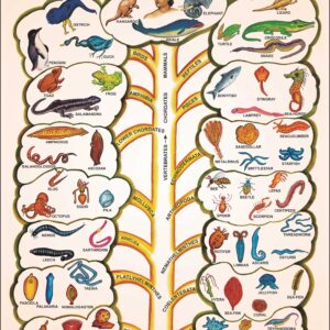
(2) Animal kingdom
(3) L.H. of frog(development)
(4) L.H. of frog(metamorphosis)
(5) L.H. of mosquito
(6) Epithelial & connective tissues
(7) Simple and complex tissues
(8) Rabbit dissection and skeletonWhile size 25 x 35 cm is available in sets only, other sizes are available singly as well.
BG13161 -
Charts
Zoology Series III
These vacuum-formed biological charts are made from heavy-duty plastic, offering deep relief for clear, tactile representation. Weatherproof and washable, they are built to withstand years of
classroom use. With realistic colors and an aesthetically pleasing design, these charts provide valuable assistance in helping students understand biological concepts. They are also specially designed to be helpful for blind students, making learning more accessible.Frog
Set of 8
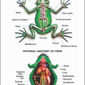
(1) Frog anatomy and skeleton
(2) Frog digestive system
(3) Frog circulatory system
(4) Frog respiratory system
(5) Frog nervous system
(6) Frog reproductive system, male
(7) Frog reproductive system, female
(8) Frog heartWhile size 25 x 35 cm is available in sets only, other sizes are available singly as well
BG13163 -
Charts
Zoology Series IV
These vacuum-formed biological charts are made from heavy-duty plastic, offering deep relief for clear, tactile representation. Weatherproof and washable, they are built to withstand years of
classroom use. With realistic colors and an aesthetically pleasing design, these charts provide valuable assistance in helping students understand biological concepts. They are also specially designed to be helpful for blind students, making learning more accessible.Rat
Set of 8
(1) Rat anatomy,dissection showing internal organs (female)
(2) Rat digestive system
(3) Rat circulatory system
(4) Rat respiratory system
(5) Rat excretory system
(6) Rat reproductive system (male)
(7) Rat reproductive system (female)
(8) Rat brain & heartWhile size 25 x 35 cm is available in sets only, other sizes are available singly as well
BG13165 -
Charts
Zoology Series V
These vacuum-formed biological charts are made from heavy-duty plastic, offering deep relief for clear, tactile representation. Weatherproof and washable, they are built to withstand years of
classroom use. With realistic colors and an aesthetically pleasing design, these charts provide valuable assistance in helping students understand biological concepts. They are also specially designed to be helpful for blind students, making learning more accessible.Set of 8
(1) Life History of Honey Bee
(2) Life History of Silkworm
(3) Life History of House Fly
(4) Malarial Parasite (Plasmodium)
(5) Cockroach, circulatory & nervous systems
(6) Cockroach, external features
(7) Cockroach, digestive & respiratory systems
(8) Earthworm, circulatory & excretory systemsWhile size 25 x 35 cm is available in sets only, other sizes are available singly as well.
BG13167 -
Charts
Human Skin
These vacuum-formed biological charts are made from heavy-duty plastic, offering deep relief for clear, tactile representation. Weatherproof and washable, they are built to withstand years of classroom use. With realistic colors and an aesthetically pleasing design, these charts provide valuable assistance in helping students understand biological concepts. They are also specially designed to be helpful for blind students, making learning more accessible.
BG13169 -
Charts
Types of Human Muscular Tissues
These vacuum-formed biological charts are made from heavy-duty plastic, offering deep relief for clear, tactile representation. Weatherproof and washable, they are built to withstand years of classroom use. With realistic colors and an aesthetically pleasing design, these charts provide valuable assistance in helping students understand biological concepts. They are also specially designed to be helpful for blind students, making learning more accessible.
BG13173 -
Charts
Human Endocrine System
These vacuum-formed biological charts are made from heavy-duty plastic, offering deep relief for clear, tactile representation. Weatherproof and washable, they are built to withstand years of classroom use. With realistic colors and an aesthetically pleasing design, these charts provide valuable assistance in helping students understand biological concepts. They are also specially designed to be helpful for blind students, making learning more accessible.
BG13175 -
Plant Physiology
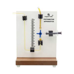
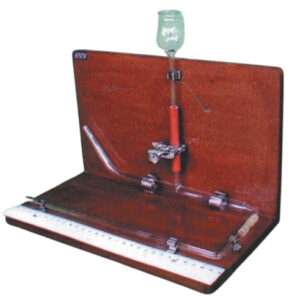
Potometer Apparatus
This Potometer Apparatus provides a simple and effective way to demonstrate plant transpiration by measuring water absorption. The transparent graduated scale allows clear observation, while the syringe and control valve make it easy to refill and regulate water flow. The complete setup is mounted on a sturdy base, making it highly suitable for laboratory demonstrations and plant physiology studies.
Features:
- Transparent graduated scale for accurate readings
- Tubing system with control valve for smooth regulation
- Syringe for convenient refilling and adjustments
- Stable base for durability and ease of handling
- Ideal for demonstrating water uptake in plants
BG13335P -
Plant Physiology
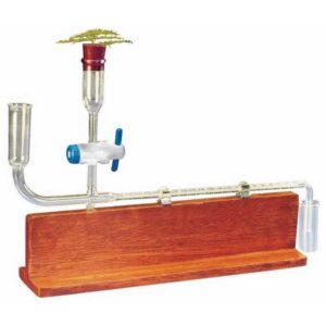
Potometer, Ganong’s (On Wooden Stand)
Designed to demonstrate the rates of transpiration and water absorption in a cut shoot, this setup includes a graduated capillary tube bent at a right angle with a small orifice. The tube is connected to a water reservoir with a glass stopcock for controlled water flow. A rubber stopper at the opposite end securely holds the plant shoot. The entire apparatus is mounted on a polished wooden stand, ensuring stability during use. Ideal for educational and laboratory demonstrations.
BG13337 -
Plant Physiology
Potometer, Ganong’s (On Plastic Stand)
This apparatus demonstrates the rates of transpiration and water absorption in a cut shoot. It features a graduated capillary tube bent at a right angle with a small orifice, connected to a water reservoir with a glass stopcock for precise control. A rubber stopper at the other end holds the plant shoot securely. The entire setup is mounted on a sturdy wooden stand for stability, making it ideal for laboratory and classroom use.
BG13339 -
Plant Physiology
Potometer, Student’s
This apparatus includes a 160 x 20 mm glass tube with an 8 mm side arm for holding a plant. It comes with a solid rubber stopper at the top and a rubber stopper with a hole at the bottom for secure fitting. A 140 mm capillary tube is also included, allowing accurate measurement of water uptake. Ideal for demonstrating plant transpiration and water absorption in laboratory experiments.
BG13340 -
Plant Physiology
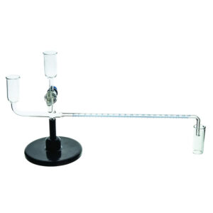
Potometer, Thoday’s
Provided in a glass tube with a diameter of 15 mm, featuring a side arm leading to a thistle funnel embedded in rubber tubing. Both ends of the glass tube are tapered and bent at right angles. One end is designed to accommodate the plant stem, while the other end is connected to a capillary tube with a milimeter scale. This entire set up is mounted on an L-shaped stand for stability and ease of use.
BG13347 -
Plant Physiology
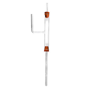
Potometer Darwin
Designed to measure water absorption in transpiring plants, this apparatus includes a glass tube with a straight limb. The upper end is sealed with a rubber stopper, while the lower end is fitted with a bored rubber stopper to accommodate a capillary tube. Ideal for studying plant water uptake in laboratory experiments.
BG13349 -
Plant Physiology
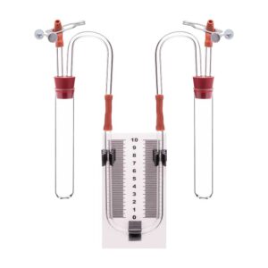
Respiroscope, Simple
This compact unit is designed for comparing the rates of gas absorption or evolution during respiration in small organisms. It includes two stoppered boiling tubes connected by three-way taps at both ends of a manometer. One tube holds the organisms and features a syringe for adjusting the liquid level, while the second tube serves as a control thermo-barometer. This setup enables
accurate measurement and comparison of gas exchange rates in laboratory experiments.BG13370