Botanical models
-
-
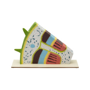
Botanical models
MONOCOT STEM INTERNAL ANATOMY & VASCULATURE
This model showcases the different tissues and vascular bundles of maize, including scattered, closed, and collateral types, in both transverse and longitudinal sections. It allows for detailed examination of internal structures such as large pitted vessels and spiral and annular vessels, revealing cell wall thickenings in longitudinal sections. Highlights parts like the cortex, endodermis, and pericycle. Mounted on a base and accompanied by a key card for reference.
BG12862 -
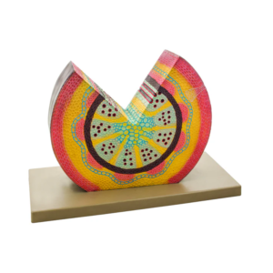
Botanical models
MODEL MONOCOT STEM
This model showcases the tissue composition, as well as the scattered closed and collateral vascular bundles, in both transverse and longitudinal sections of maize. Highlighting features such as large pitted vessels, spiral, and annular vessels. Mounted on a base and accompanied by an English Key Card.
BG12862A -
Botanical models
DICOT STEM ANATOMY MODEL- TRANSVERSE AND LONGITUDINAL SECTIONS
This anatomical model displays both transverse (T.S.) and longitudinal (L.S.) sections of a dicotyledonous stem, emphasizing the structure at the stage where the cambium ring has formed but secondary growth has not yet occurred. It’s designed for studying the intricate anatomy of dicot stems, featuring key components such as the epidermis, lenticels, cork layer, cork cambium, cortical parenchyma, starch sheath, medullary rays, phloem (including sieve plates, sieve tubes, and phloem parenchyma), interfascicular cambium, xylem (with pitted vessels, bordered pitted vessels, annular vessels, and spiral vessels), and pith. Mounted on a stable base, the model includes a key card that labels and explains each part, making it an invaluable educational tool for students, educators, and researchers exploring plant anatomy and biology.
BG12867B -
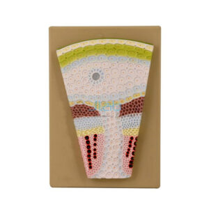
Botanical models
MONOCOT STEM T. S.
Explore the internal structure of a monocot stem from maize with this model, featuring transverse sections of various tissues and vascular bundles. Each section is comprised of a single layer of cells with a thick cuticle. Stomata are present, while multicellular hairs are absent. Mounted on a board for display, accompanied by a key card for easy reference.
BG12872 -
Botanical models
LEAF ANATOMY
Intricate structure of a leaf with this 3-dimensional model, showcasing detailed transverse and longitudinal sections. Featuring loosely packed spongy mesophyll and stomata beneath for gas exchange, this large-size model is mounted on a stand for easy viewing. Accompanied by a key card for reference.
BG12880 -
-
Botanical models
CHLOROPLAST MODEL
This model provides a comprehensive exploration of key chloroplast features, including ribosomes, DNA, starch granules, membranes, stroma, thylakoids, granum, lamellae, and lumen. Each feature is colored and numbered for easy identification, with an included key card for reference.
BG12990 -
Botanical models
PLANT CELL ANATOMY STUDY MODEL
Featuring key structures like the cell wall, plasma membrane, nucleus, chloroplasts, and more, it’s ideal for studying plant cell anatomy. Each feature is numbered and color-coded for easy identification, with an included key card for reference. This freestanding model offers an effective method for exploring the intricate structures and functions within a plant cell.
BG12992